
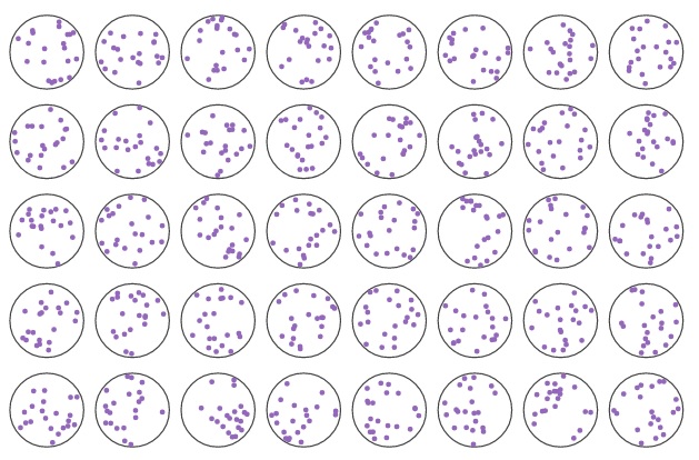
Random spread of 21 teoretical cells in the LCMD area.

|
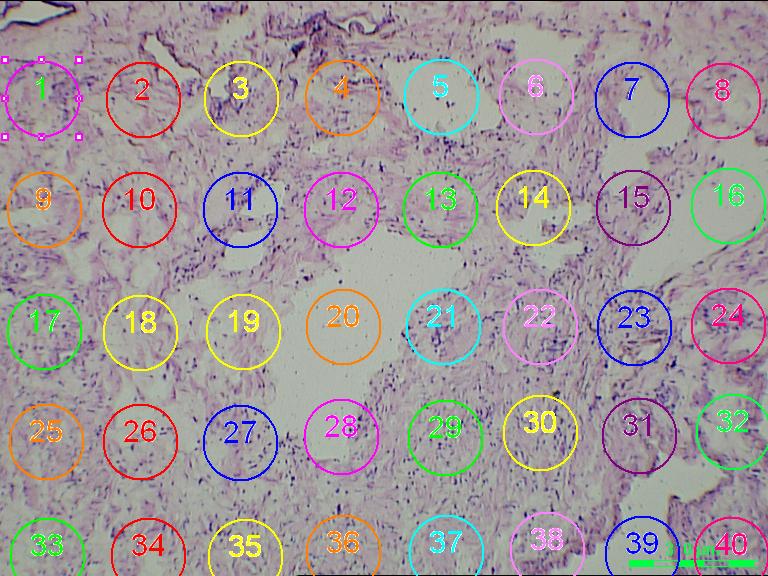
Picture of 40 circle that is part of the 96 grid used in the LCMD procedure. Dark purple (dark blue to violet) spots are nuclei stain by Giemsa. The picture below demonstrates a non-uniform distribution of the nuclei within each LCMD cut.
|
||||||||||||||||||
 |
||||||||||||||||||
|
The theoretical spread of 21 cell either homoplasmic mutant or pure wild-type. Cells are given the value 1 if the mutant fraction is large than zero, and 0 when the mutant fraction becomes less than zero. Starting with the mutant fraction (in an LCMD cut) the first cell is given the value 1, then subtracting 5% point from the mutant fraction and checking if the value is above or below zero and reassigning a new value for the second cell. This iteration is performed 21 times for each mutant fraction. For example, a mutant fraction of 11% will result in 3 cells with value 1 and 18 cells with value 0. A mutant fraction of 100% will result in 21 cells with the value 1. Likewise, a mutant fraction of 0% will result in 21 cells with the value 0 (non-mutant). The cell coordinates (X, Y, Z) and cell values are the underlying data for the isosurfaces generated on the "spread data". The figure below shows the random spread of 21 cell within an LCMD area.
Excel file with all the mutant fractions and X Y Z coordinates.
Isosurfaces depict either mutated or non-mutated cell distribution within the sample volume. The sampling volume was X = 1.5mm, Y = 2.2mm, and Z depending on the tissue thickness, ranging from 0.1mm up to 0.8mm.
| #5 Testicular tumor (Leydig cell tumor) | ||
| Marker 1 (P2F1) | Non-mutated | Mutated |
| Marker 2 (P2F2) | Non-mutated | Mutated |
| #13 Liver tumor (Hepatocellular carcinoma) | ||
| Marker 1 (P1F15) | Non-mutated | Mutated |
| #29 Colon tumor (Colon cancer) | ||
| Marker 1 (P2F1) | Non-mutated | Mutated |
| #170 Breast tumor (Breast cancer) | ||
| Marker 1 (P1F1) | Non-mutated | Mutated |
| #170 Lymph node (from breast cancer patient) | ||
| Marker 1 (P1F1) | Non-mutated | Mutated |
| #170 Liver metastases (from breast cancer patient) | ||
| Marker 1 (P1F1) | Non-mutated | Mutated |
| # 231 Bladder tumor (Bladder cancer) | ||
| Marker 1 (P2F6) | Non-mutated | Mutated |
| Marker 2 (P5F6) | Non-mutated | Mutated |
| #231 Lymph node, (from bladder cancer patient) | ||
| Marker 1 (P2F6) | Non-mutated | Mutated |
| Marker 2 (P5F6) | Non-mutated | Mutated |