
Isosurface of mitochondrial mutation in cells within 3D tumor volumes
Most of the figures and pictures can be enlarged or interacted
|
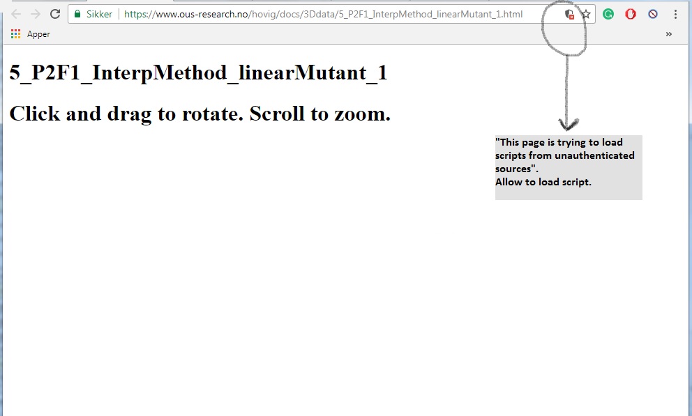
If the view of 3D isosurfaces is not displayed, click on the shield symbol in the URL "box" and overwrite "This page is trying to load scripts from unauthenticated sources". Link to picture script activation. Or deactivate the secure web browsing mode in the web browser. |
Alternatively, data is plotted in Plotly.com (Link to Plotly graphs.) |
50% mutant fraction in mtDNA |
96 laser capture microdissection (LCMD) samples were taken from numerous 12-micrometer tumor slices. Each sample was analyzed for known somatic mtDNA mutation. Some of the samples had two mtDNA mutations, thus both were analyzed in the LCMD samples. A total of 9504 laser capture microdissection samples was collected from these tissues.
Based on the mutant fraction found in each LCMD sample a random distribution of 21 cells (mutated and non-mutated) was given X, Y, Z coordinates in the LCMD sample volume. For detailed description follow this LINK.
Isosurfaces depict either mutated or non-mutated cell distribution within the sample volume. The sampling volume was X = 1.5mm, Y = 2.2mm, and Z depending on the tissue thickness, ranging from 0.1mm up to 0.8mm. Illustration of z-axis sampling and tissue support (membrane or glass slide) are shown in this LINK.
Data: Raw data in an Excel format can be downloaded here.
Tissue analyzed
| #5 Testicular tumor (Leydig cell tumor) | ||
| Marker 1 (P2F1) | Non-mutated | Mutated |
| Marker 2 (P2F2) | Non-mutated | Mutated |
| #13 Liver tumor (Hepatocellular carcinoma) | ||
| Marker 1 (P1F15) | Non-mutated | Mutated |
| #29 Colon tumor (Colon cancer) | ||
| Marker 1 (P2F1) | Non-mutated | Mutated |
| #170 Breast tumor (Breast cancer) | ||
| Marker 1 (P1F1) | Non-mutated | Mutated |
| #170 Lymph node (from breast cancer patient) | ||
| Marker 1 (P1F1) | Non-mutated | Mutated |
| #170 Liver metastases (from breast cancer patient) | ||
| Marker 1 (P1F1) | Non-mutated | Mutated |
| # 231 Bladder tumor (Bladder cancer) | ||
| Marker 1 (P2F6) | Non-mutated | Mutated |
| Marker 2 (P5F6) | Non-mutated | Mutated |
| #231 Lymph node, (from bladder cancer patient) | ||
| Marker 1 (P2F6) | Non-mutated | Mutated |
| Marker 2 (P5F6) | Non-mutated | Mutated |
Alternative display:
An alternative way of presenting these data is by creating isosurfaces for the mutant fractions found in each LCMD sample. As an example, all isosurfaces of mutant fractions from 0% and up to 100% (in 5% point increments) can be found for the samples #5 testicular cancer and #231 lymph node (bladder cancer). Please note that when the isosurface is created for each mutant fraction the intermediate mutant fractions are not part of the interpolation. Furthermore, a lower resolution was also used as compared to the "spread" of 21 theoretical cells in the LCMD volume.
Isosurface's was created in Matlab with the following script:
tx=90:18:1350
ty=90:18:2070
tz=-564:6:0
[xg,yg,zg] = meshgrid(tx,ty,tz);
F.Method = 'natural';
F = TriScatteredInterp(x, y, z, e);
eg = F(xg,yg,zg);
fv=isosurface(xg,yg,zg,eg,MF);
MF=[0%, 5%. . . . . . . . . . . . . . . . 100%]
|
The Norwegian Radium hospital Paulo Refinetti Stephan Morgenthaler William Thilly Per Olaf Ekstrøm Paulo Refinetti Stephan Morgenthaler William Thilly Per Olaf Ekstrøm Paulo Refinetti Stephan Morgenthaler William Thilly Per Olaf Ekstrøm Paulo Refinetti Stephan Morgenthaler William Thilly Per Olaf Ekstrøm Paulo Refinetti Stephan Morgenthaler William Thilly Per Olaf Ekstrøm Paulo Refinetti Stephan Morgenthaler William Thilly Per Olaf Ekstrøm Paulo Refinetti Stephan Morgenthaler William Thilly Per Olaf Ekstrøm Paulo Refinetti Stephan Morgenthaler William Thilly Per Olaf Ekstrøm Paulo Refinetti Stephan Morgenthaler William Thilly Per Olaf Ekstrøm Paulo Refinetti Stephan Morgenthaler William Thilly Per Olaf Ekstrøm Paulo Refinetti Stephan Morgenthaler William Thilly Per Olaf Ekstrøm Paulo Refinetti Stephan Morgenthaler William Thilly Per Olaf Ekstrøm Paulo Refinetti Stephan Morgenthaler William Thilly Per Olaf Ekstrøm Cycling temperature capillary electrophoresis MegaBACE mtDNA mutation analysis Cycling temperature capillary electrophoresis MegaBACE mtDNA mutation analysis Cycling temperature capillary electrophoresis MegaBACE mtDNA mutation analysis Cycling temperature capillary electrophoresis MegaBACE mtDNA mutation analysis Cycling temperature capillary electrophoresis MegaBACE mtDNA mutation analysis Cycling temperature capillary electrophoresis MegaBACE mtDNA mutation analysis Cycling temperature capillary electrophoresis MegaBACE mtDNA mutation analysis Cycling temperature capillary electrophoresis MegaBACE mtDNA mutation analysis Cycling temperature capillary electrophoresis MegaBACE mtDNA mutation analysis Cycling temperature capillary electrophoresis MegaBACE mtDNA mutation analysis Cycling temperature capillary electrophoresis MegaBACE mtDNA mutation analysis Cycling temperature capillary electrophoresis MegaBACE mtDNA mutation analysis Cycling temperature capillary electrophoresis MegaBACE mtDNA mutation analysis |