Sample #231 Bladder tumor

Overview of tissue
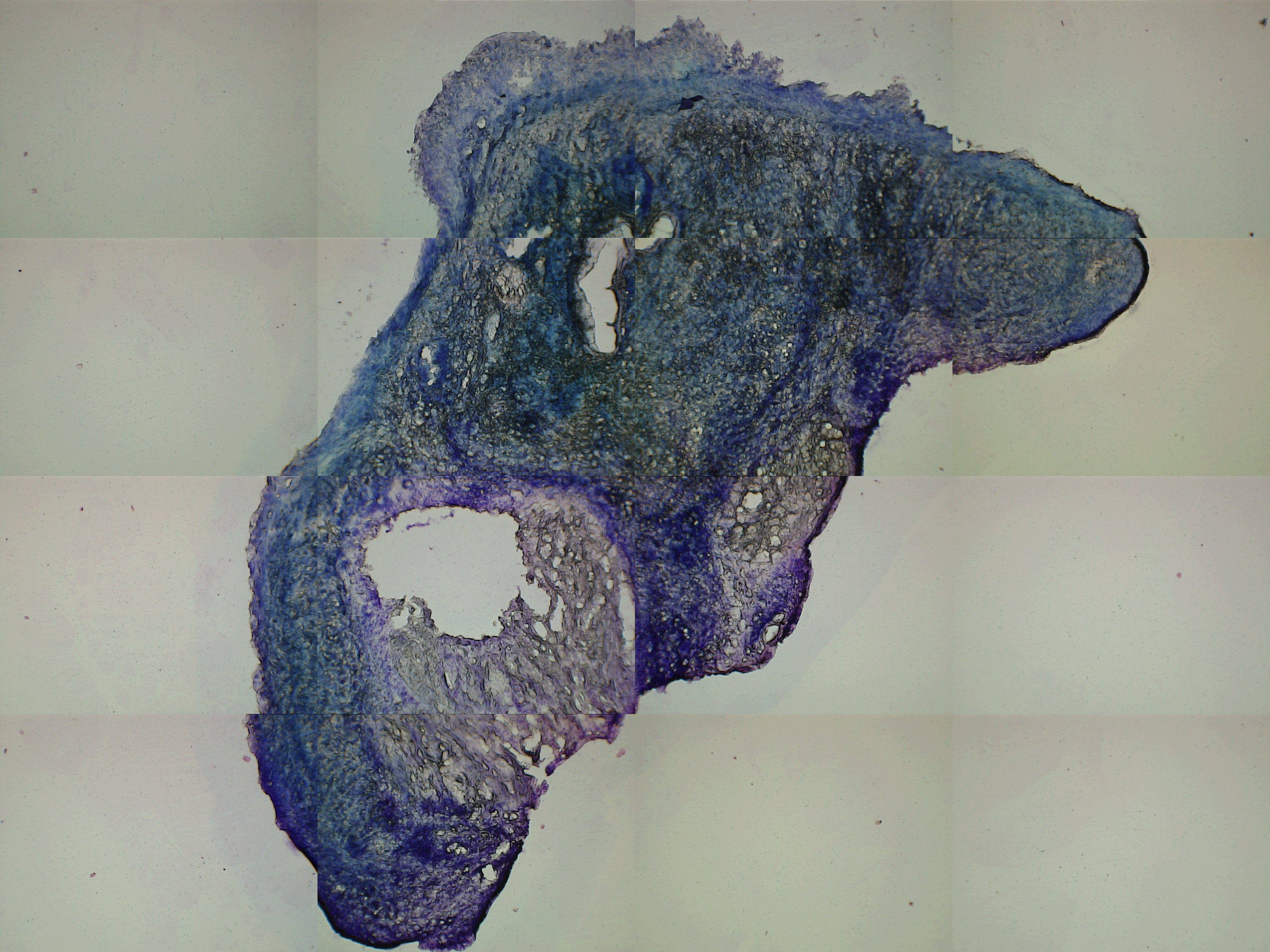
This sample was a tiny tissue piece that had dried in the -70°C freezer. Attempts were made to rehydrate the tissue prior to sectioning in the cryotome. The reason for this deviation from the standard protocol was that a matching lymph node (in good condition) was available. Consequently thicker slices had to be taken. 9 microtome slices, of 20 μm thickness were obtained, where 4 slices were subjected to laser capture microdissection (LCMD). The same grid of 96 (~25000 μm2) circles was used as a template for the laser capture microdissection on each slide.
Descriptive statistics of mutant fraction found in each LCMD sample.
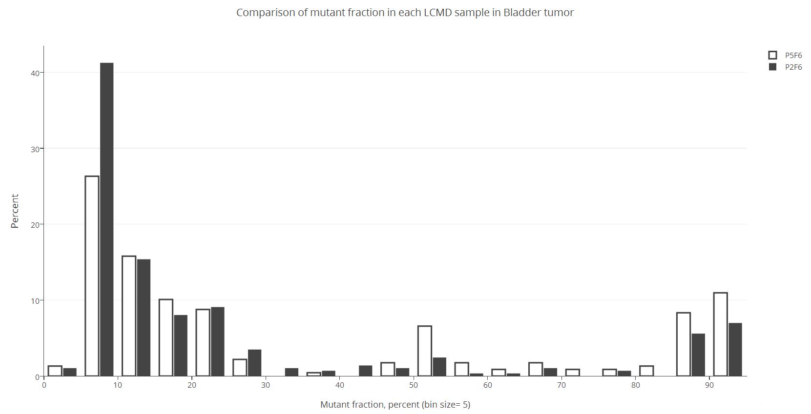
| P2F6 (M1) |
P5F6 (M2) | |
| N | 286 | 228 |
| Min (%) | 1.5 | 4.4 |
| Max (%) | 93.8 | 93.7 |
| Mean (%) | 25.5 | 35.1 |
| First Quantile (%) | 8.24 | 9.7 |
| Median (%) | 11.5 | 18.7 |
| Third Quantile (%) | 25.0 | 59.3 |
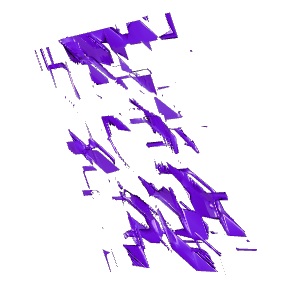
Links to 3 different approaches to visualizing the mutant fraction in the 3D volume.
Isosurface of a random spread of homoplasmic cells (wild-type and/or mutant) in each LCMD volumes.
| P2F6 (M1) | Non-mutated | Mutated |
| P5F6 (M2) | Non-mutated | Mutated |
No isosurfaces of mutant fractions were created for the two markers due condition of the tissue and the sampling.
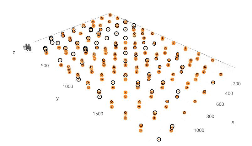
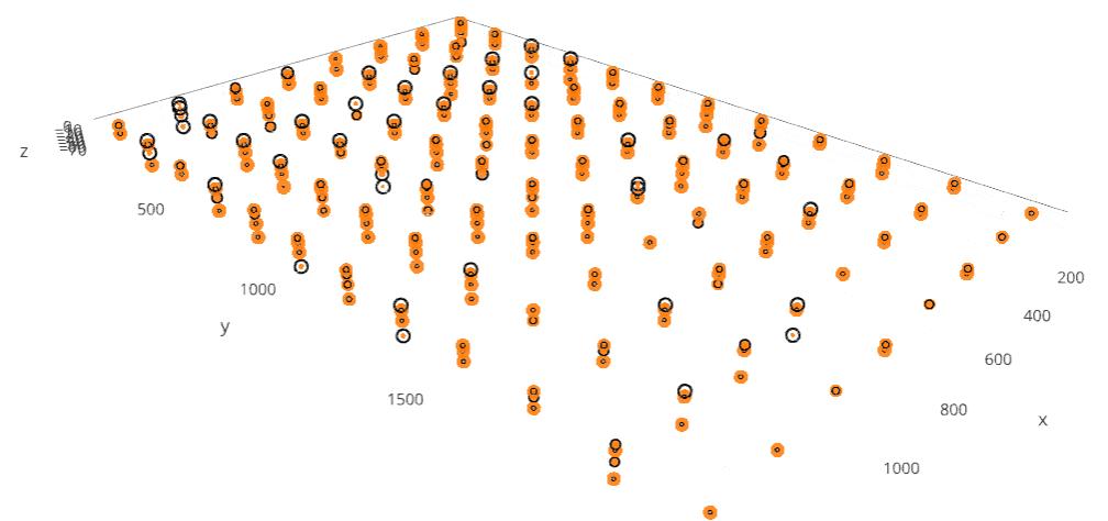
3D bubble plot of wild-type and mutant fractions.
Marker 1 (P2F6)
Marker 2 (P5F6)