
Cancer covers a broad spectrum of diseases, in every tissue of the body. Tissues are composed of cells, which normally grow slowly, under the tight control of a network of regulatory genes. The slow accumulation of activating mutations in growth genes, and inactivating mutations in suppressor genes, eventually allows a cell to grow out of control. Relapse is due to the development of resistant cells, rather than the escape of sensitive cells, suggesting the need for new approaches to treatment of the disease.
Peptide nucleic acids
In this project we use modified peptide nucleic acids (PNA) combined with photochemical internalisation (PCI) technology. We are working on following topics:
- Developing a treatment combination to easily knock out any gene of interest in an experimental system.
- Systematic screening of gene products after knocking out a specific gene of interest.
- Validate uptake, transport and localisation of PNA linked to different peptide signals.
We are using molecular tools like laser-induced capillary electrophoresis, fluoroscens- and confocal microscopy, flow cytometry, photochemical internalisation technology and microarray analysis.
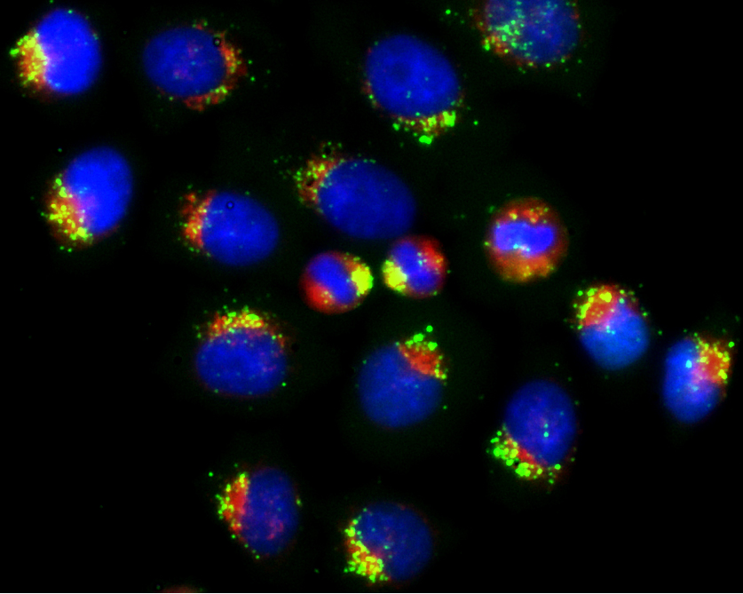
Demonstration of the PNA/PCI principle in living cells
To view the distribution of PNA molecules without PCI, click here
To view the distribution of PNA molecules with PCI, click here