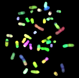
Fluorescent in situ hybridization (FISH) is a powerful technique that uses non-toxic fluorescent DNA probes to target any given sequence within a nucleus, resulting in colored signals that are detected with a fluorescence microscope. It circumvents the need for tumor cell-culture (fresh or paraffin-embedded interphase nuclei can be analyzed directly), and provides researchers with a quick and precise screening method over large quantities of cells.
Standard FISH techniques with chromosome or locus-specific probes can identify rearrangements too subtle or too complex to be disclosed by classical chromosome banding, with the downside of revealing only those aberrations one tests for. This is therefore not suitable for the initial screening of samples with an unknown genetic background. However, recent developments have made available FISH-based techniques with much wider specificity than the original method, such as multicolor-FISH (M-FISH) and comparative genomic hybridization (CGH), which our group has extensive experience in performing.
More specifically, M-FISH uses a pool of whole-chromosome painting probes with different fluorochrome combinations that result in specific color patterns for each human chromosome. It can detect complex inter-chromosomal rearrangements or changes involving segments too small or too similarly banded to be adequately described by standard banding, with the downside of requiring metaphases (i.e., tumor cell culture) to be performed.
M-FISH metaphase spread of colon cancer cell line V9P (Kleivi et al. (2004))
Method overview
Interphase FISH (IP-FISH). For fresh samples, tissue is disaggregated (with scalpel and/or enzymatic solution) and the isolated cells are treated with a hypotonic solution (to burst the plasma membranes and free the nuclei), fixed in methanol and dropped onto glass slides. A quick pre-treatment procedure is performed using a warm solution of 2xSSC (to remove cytoplasm background), afterwhich slides are dehydrated in an alcohol series and placed in an automated denaturation/hybridization system. The selected probes, labeled with different fluorochromes, are added to each slide at this step, and a co-denaturation procedure is then performed by raising the temperature to 72-75 ºC for 2-5 minutes, followed by a 16-24 h hybridization period at 37 ºC. Finally, the excess probe is washed off the slides, a DNA counterstain is added (usually DAPI), and the cells are then analyzed under a fluorescence microscope. For paraffin-embedded material, several additional steps are required: the paraffin needs to be melted and removed using xilol and ethanol, and a pre-treatment procedure with a high-salt solution (at high temperature) is recommended, to facilitate probe entrance into the nuclei. An enzymatic digestion step is also required, as most paraffin sections contain whole cells rather than isolated nuclei, and the cytoplasmic content would affect hybridization. The rest of the procedure is similar to the one described for fresh tissue, but extra care should be taken not to lose the thin tissue sections during the washing steps.
Multicolor FISH (M-FISH) Fresh samples need to be cultured and processed as for normal karyotyping. A glass slide containing metaphase cells for the sample is then obtained, pre-treated in 2xSSC and usually subjected to an enzymatic treatment (pepsin) to facilitate probe entrance. Co-denaturation does not perform well for M-FISH, so two separate denaturation steps need to be carried out: on the one side, the pre-treated metaphase slides are dehydrated and placed on a formamide solution at 72-75 ºC for a few minutes. On the other side, commercially available M-FISH probe cocktails, labeled and combined to produce a unique color for each chromosome, are denaturated in a heating plate according to the manufacturers instructions. The probe mixture is then added to the slides, and hybridization is allowed to occur for 24-72 h at 37 ºC, followed by stringency washes. Slides are then counterstained and fluorescent images corresponding to the different fluorochromes are sequentially captured and merged using software specifically designed for this purpose.
Totland MZ, Knudsen LM, Rasmussen NL, Omori Y, Sørensen V, Elster VCW, Stenersen JM, Larsen M, Jensen CL, Zickfeldt Lade AA, Bruusgaard E, Basing S, Kryeziu K, Brech A, Aasen T, Lothe RA, Leithe E(2024) The E3 ubiquitin ligase ITCH negatively regulates intercellular communication via gap junctions by targeting connexin43 for lysosomal degradation Cell Mol Life Sci, 81(1), 171 DOI 10.1007/s00018-024-05165-8, PubMed 38597989
Majid U, Bergsland CH, Sveen A, Bruun J, Eilertsen IA, Bækkevold ES, Nesbakken A, Yaqub S, Jahnsen FL, Lothe RA(2024) The prognostic effect of tumor-associated macrophages in stage I-III colorectal cancer depends on T cell infiltration Cell Oncol (Dordr)(in press) DOI 10.1007/s13402-024-00926-w, PubMed 38407700
Sveen A, Johannessen B, Klokkerud SM, Kraggerud SM, Meza-Zepeda LA, Bjørnslett M, Bischof K, Myklebost O, Taskén K, Skotheim RI, Dørum A, Davidson B, Lothe RA(2024) Evolutionary mode and timing of dissemination of high-grade serous carcinomas JCI Insight, 9(3) DOI 10.1172/jci.insight.170423, PubMed 38175731