Coordination of cell division and cell migration

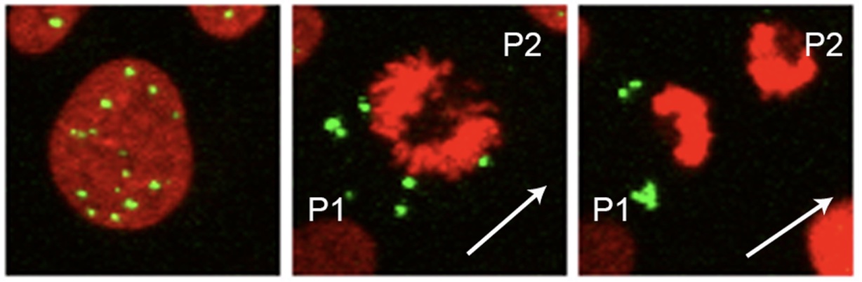
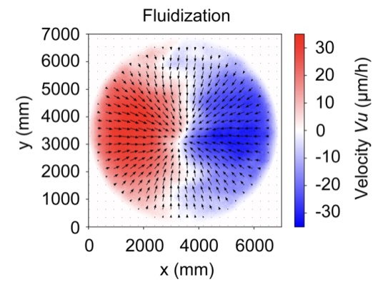
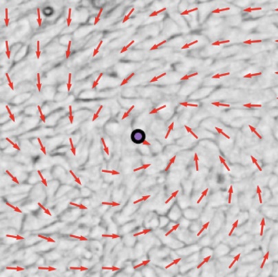
Cells possess the ability to both divide and migrate concurrently during instances of wound healing, development, and cancer progression. But how are these two processes coordinated? We have shown that epithelial cells divide asymmetrically along the axes of cell sheet displacement. These cell divisions are characterized by generation of an asymmetric chromatin configuration, leading to the uneven segregation of organelles, such as PML bodies and lysosomes, to daughter cells.

Link to paper: 
Coordinated collective migration and asymmetric cell division in confluent human keratinocytes without wounding.
Lång E, Połeć A, Lång A, Valk M, Blicher P, Rowe AD, Tønseth KA, Jackson CJ, Utheim TP, Janssen LMC, Eriksson J, Bøe SO.
Nat Commun. 2018 Sep 10;9(1):3665. doi: 10.1038/s41467-018-05578-7.
PMID: 30202009


Mechanical forces and tissue dynamics during transition from quiescence to proliferation.

The majority of cells within the human body remain dormant, exhibiting slow growth and limited motility. However, in processes like wound healing, stem cell activation, immune cell activation, and cancer development, cells transit to a more active state marked by increased proliferation and migration. Our research has unveiled and comprehensively analyzed a novel mechanism governing the movement of epithelial cell sheets after exit from quiescence. This mechanism is intricately linked to the activation of dormant mechanical forces inherent in quiescent epithelial monolayers.

Link to paper: 
Mechanical coupling of supracellular stress amplification and tissue fluidization during exit from quiescence.
Lång E, Pedersen C, Lång A, Blicher P, Klungland A, Carlson A, Bøe SO.
Proc Natl Acad Sci U S A. 2022 Aug 9;119(32):e2201328119. doi: 10.1073/pnas.2201328119. Epub 2022 Aug 1.
PMID: 35914175


Polar self-organization of dynamic epithelial monolayers.

The capacity of epithelial monolayers to autonomously adopt a dynamic polarized state plays a crucial role in tissue regeneration, developmental processes, and tumor progression. We have developed a novel experimental approach aimed at investigating the self-organizing potential of epithelial monolayers. In these experiments cell monolayers undergo a transition from a disordered and static state in quiescence to a dynamic state characterized by extensive long-range polar alignment when exposed to growth factors. Our research reveals that this self-organizing phenomenon hinges on the formation and subsequent dissolution of dynamic topological defects, which carry +1 and -1 charges.

Link to paper:

Lång E, Lång A, Blicher P, Rognes T, Dommersnes PG, Bøe SO (2024)
Topology-guided polar ordering of collective cell migration
Sci Adv, 10 (16), eadk4825
DOI 10.1126/sciadv.adk4825, PubMed 38630812


Advanced Light Microscopy

The research group oversees the advanced light microscopy core facility node situated at Rikshospitalet. The facility has been established in partnership with the South-Eastern Norway Regional Health Authority, Oslo University Hospital and the University of Oslo. It features a diverse range of cutting-edge microscope technologies, including live cell imaging, high-content imaging, confocal microscopy, and super-resolution microscopy. The facility provides comprehensive hands-on training in advanced microscopy techniques and offers support for processing and analysis of imaging data.

Link to facility home page