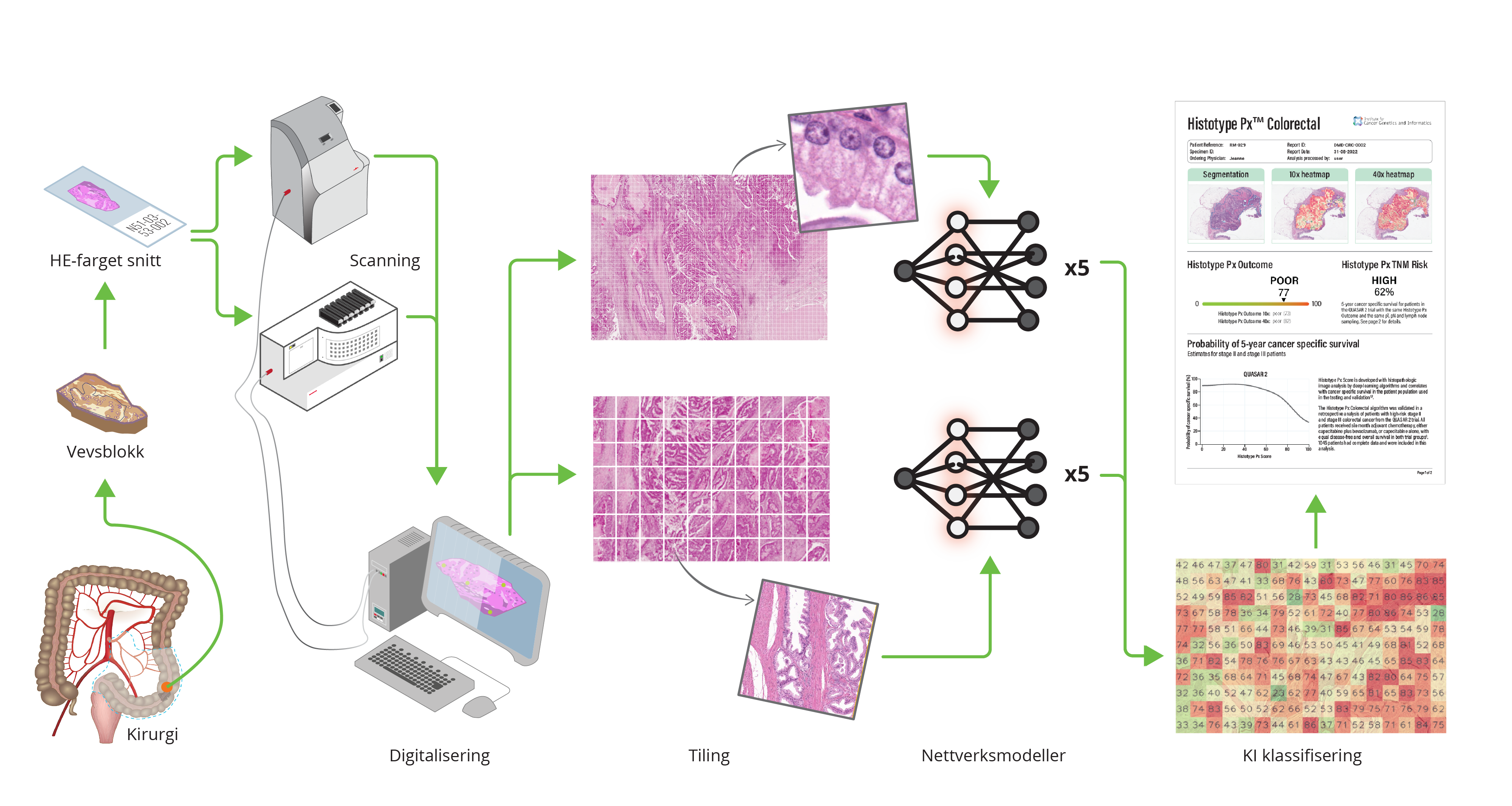
One of the products emerging from the DoMore! project is called “Histotyping”, which uses the marker DoMore-v1-CRC, and culminated in 2020 in a landmark artificial intelligence (AI) publication in the Lancet (Skrede et al., The Lancet 2020).
Histotyping involves an automated analysis of high-resolution scans of cancerous tissue stained with routine hematoxylin and eosin (H&E). ICGI developed convolutional neural networks for this method and trained on extensive retrospective clinical trial data with known patient outcomes. Deep learning models were trained to identify and analyse the morphology of a tumour in a surgically resected tissue section. Histotyping estimates the probability of a good or poor outcome, i.e., the likelihood of a recurrence. It could be employed as a supplementary diagnostic service to support clinicians and patients in their adjuvant chemotherapy decision-making for patients with stages II and III CRC.
Histotyping as explained for the Norwegian TV channel TV2.
The DoMore-v1-CRC marker has also been combined with established clinical pathology markers (T and N stage, and number of harvested lymph nodes) in a decision tree solution for clinical decision support. Compared to conventional risk stratification for adjuvant therapy, the proposed method identified a significantly larger group of patients with a good prognosis that are candidates to be spared adjuvant treatment (Kleppe et al., Lancet Oncology 2022)
While an increasing number of publications on deep learning systems claim performance comparable with or better than clinicians, few have yet demonstrated real-world medical utility. We published a Perspective in Nature Reviews Cancer in 2021 to discuss the reasons for the moderate progress and to present our recommendations for how to perform pivotal validations of deep learning systems that will provide realistic performance estimates of the systems and thereby better guide further research and clinical implementation of truly promising systems (Kleppe et al., Nat Rev Cancer 2021).
Using the DoMore-v1 network, a similar approach as for colorectal cancer has been applied to prostate cancer. The adaption of this method from CRC to PCa included using sections from multiple tissue blocks as input images, as prostate tumours are far more heterogeneous than colorectal tumours. The resulting marker, the DoMore-v1-PCa marker, predicts the risk of recurrence after radical prostatectomy and provides prognostic information supplementary to the clinical and pathological markers currently used in clinical practice. A manuscript reporting on the pivotal validation of this marker has been submitted for publication.
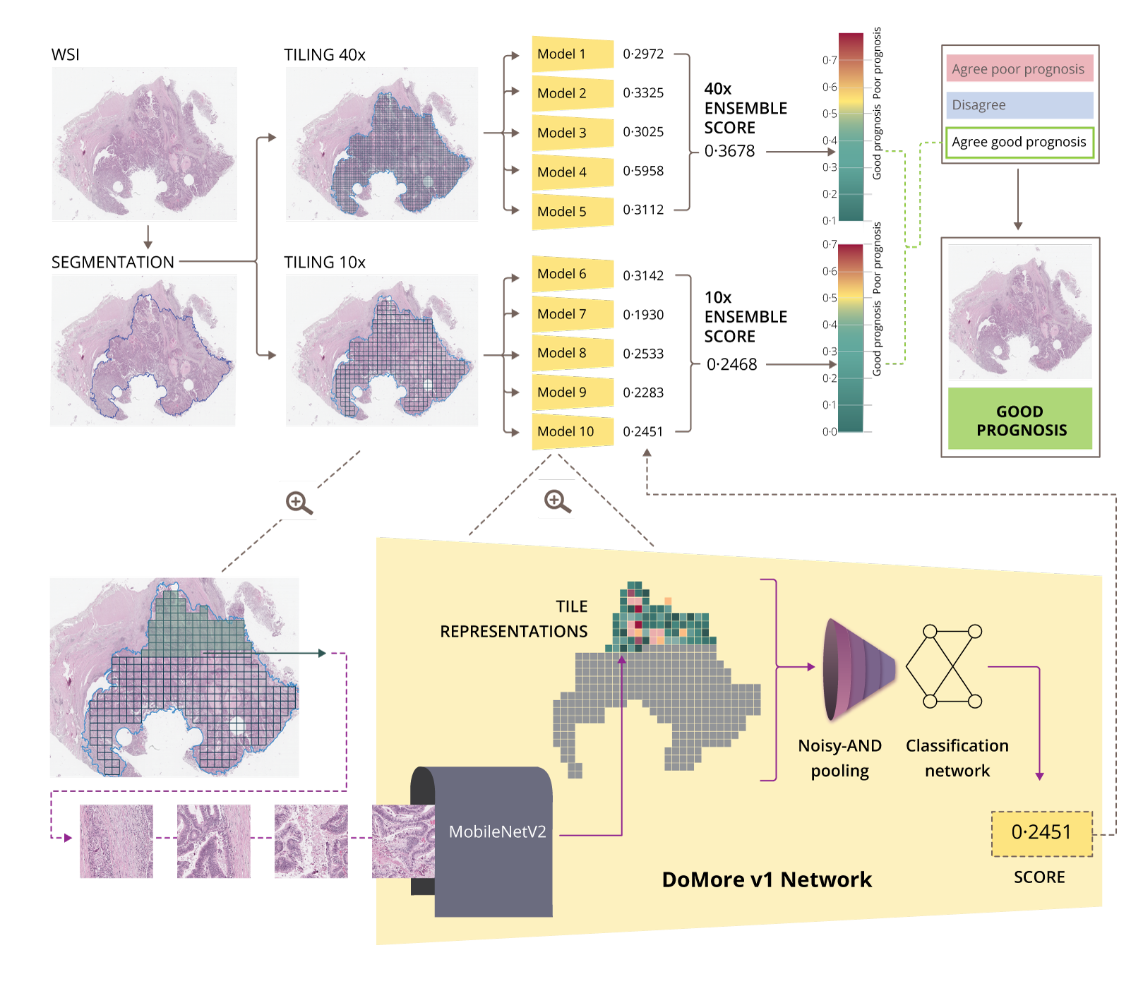
The illustration above shows the set-up of the DoMore v1 network. First the Whole Slide Image (WSI) is scanned and the tumor area segmented. Each scan is analysed 5 times at 10x resolution, and 5 times at 40x resolution. The image is split into tiles, which each are analysed in the DoMore v1 Network, giving a result relating to a a good or bad prognosis. If the number is closer to "1", the probability of a bad prognosis is larger, while as a result closer to "0" may predict a good prognosis. A patient prognosis is estimated based on the individual tiles’ prediction values. In this example both resolutions agree on a good prognosis.
Kasius JC, Kildal W, Vrede SW, Askautrud HA, Pradhan M, van Altena AM, Tobin KR, Knoll K, Reijnen C, Reimann S, Dackus G, Vlatkovic L, Huvila J, Steinlechner M, Tubita V, Gil-Moreno A, Snijders MPLM, Vos MC, Hveem TS, Asberger J, Zeimet AG, Matias-Guiu X, Lindemann K, Weinberger V, Visser NCMet al.(2025) Surgical stage in the era of molecular profiling of endometrial cancer Eur J Cancer, 233, 116164 DOI 10.1016/j.ejca.2025.116164, PubMed 41385938
Unal B, Saatcioglu F(2025) Targeting the unfolded protein response for cancer therapy: mitigating tumor adaptation and immune suppression Biomark Res, 13(1), 156 DOI 10.1186/s40364-025-00813-y, PubMed 41372991
Emblem KE, Ringstad G, Svensson SF, Vatnehol SAS, Storås TH, Vik-Mo EO, Heggebø LC, Brandal P, Eide PK(2025) Intrathecal contrast-enhanced MRI reveals an alternative route for drug delivery and brain tumor visualization Cancer Lett, 638, 218152 DOI 10.1016/j.canlet.2025.218152, PubMed 41276108